Microscopic anatomy of skeletal muscle worksheet answers – Delving into the microscopic anatomy of skeletal muscle, we embark on an exploration of the intricate structure and remarkable function of this fundamental tissue. From the diverse muscle fiber types to the hierarchical organization of muscle components, this article provides a comprehensive understanding of the skeletal muscle’s remarkable capabilities.
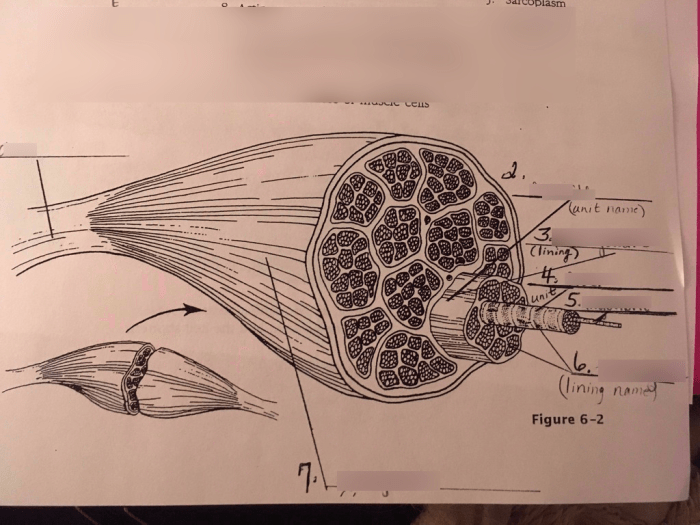
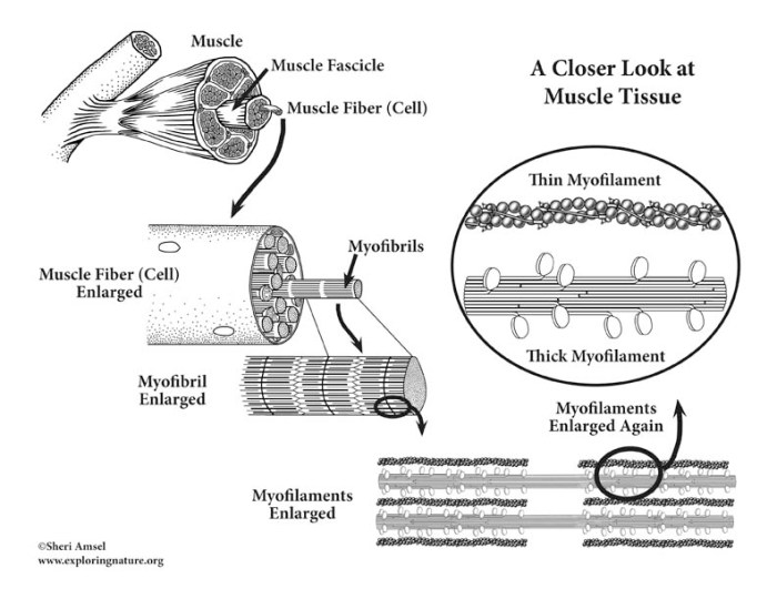
Unveiling the intricacies of muscle structure, we delve into the hierarchical organization, from the entire muscle to the individual myofibrils. The sarcomere, the fundamental unit of muscle contraction, is examined in detail, revealing the precise arrangement of thick and thin filaments that orchestrate movement.
Microscopic Anatomy of Skeletal Muscle

Skeletal muscle is a highly specialized tissue responsible for movement in the body. Understanding its microscopic anatomy is crucial for comprehending muscle function and adaptation to various stimuli.
Muscle Fiber Types
Skeletal muscle consists of different types of muscle fibers, each with distinct characteristics and functions:
- Type I (Slow-twitch) Fibers:Slow to contract and fatigue-resistant, used for endurance activities.
- Type IIa (Fast-twitch Oxidative-glycolytic) Fibers:Intermediate in speed and fatigue resistance, involved in both endurance and power activities.
- Type IIb (Fast-twitch Glycolytic) Fibers:Rapidly contracting and easily fatigued, primarily responsible for power and strength activities.
| Characteristic | Type I | Type IIa | Type IIb |
|---|---|---|---|
| Contraction Speed | Slow | Intermediate | Fast |
| Fatigue Resistance | High | Intermediate | Low |
| Primary Energy Source | Oxidative Phosphorylation | Oxidative Phosphorylation and Glycolysis | Glycolysis |
Muscle Structure
Skeletal muscle is organized hierarchically:
- Whole Muscle:A bundle of muscle fibers.
- Fascicle:A group of muscle fibers enclosed by connective tissue.
- Muscle Fiber:A single, elongated cell containing myofibrils.
- Myofibril:A bundle of thick and thin filaments responsible for muscle contraction.
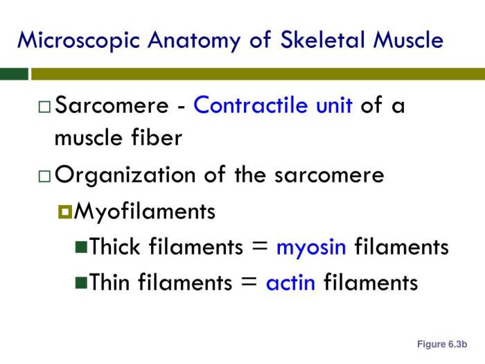
- Sarcomere:The basic unit of muscle contraction, composed of alternating bands of thick and thin filaments.
Within the sarcomere, thick filaments are made of myosin, while thin filaments are made of actin. The arrangement of these filaments creates the characteristic banding pattern seen in muscle tissue.
| Component | Description |
|---|---|
| A-Band | Region containing thick filaments |
| I-Band | Region containing only thin filaments |
| H-Zone | Central portion of the A-band where only thick filaments are present |
| M-Line | Structure in the middle of the H-zone that connects thick filaments |
| Z-Line | Structure at the edges of the I-band that anchors thin filaments |
Muscle Contraction
Muscle contraction occurs through the sliding filament theory:
- When a nerve impulse reaches a muscle fiber, it triggers the release of calcium ions from the sarcoplasmic reticulum.
- Calcium ions bind to troponin, which changes the shape of the troponin-tropomyosin complex.
- This allows myosin heads to bind to actin filaments, forming cross-bridges.
- Myosin heads then undergo a power stroke, pulling the actin filaments toward the center of the sarcomere.
- This sliding action causes the sarcomere to shorten, resulting in muscle contraction.
Muscle Metabolism, Microscopic anatomy of skeletal muscle worksheet answers
Muscle contraction requires energy, which is provided by several metabolic pathways:
- ATP (Adenosine Triphosphate):The immediate source of energy for muscle contraction.
- Creatine Phosphate:A high-energy molecule that rapidly regenerates ATP.
- Glycolysis:The breakdown of glucose to produce ATP.
- Oxidative Phosphorylation:The most efficient energy-producing pathway, utilizing oxygen to generate ATP.
| Pathway | Substrate | Products |
|---|---|---|
| ATP | N/A | ADP + Pi |
| Creatine Phosphate | Creatine Phosphate | Creatine + ATP |
| Glycolysis | Glucose | Pyruvate + ATP + NADH |
| Oxidative Phosphorylation | Acetyl-CoA | ATP + CO2 + H2O |
Muscle Adaptations
Skeletal muscle adapts to different types of exercise and training:
- Endurance Training:Increases the number and size of mitochondria, improves blood flow, and enhances the oxidative capacity of muscle fibers.
- Strength Training:Increases the cross-sectional area of muscle fibers, especially Type II fibers, and improves the recruitment of motor units.
- Plyometric Training:Enhances the elasticity and power output of muscle fibers, improving performance in activities requiring rapid force production.
Specific exercises that can improve muscle performance include:
- Endurance:Running, cycling, swimming
- Strength:Weightlifting, bodyweight exercises
- Power:Plyometrics, jumping exercises
Quick FAQs: Microscopic Anatomy Of Skeletal Muscle Worksheet Answers
What are the different types of muscle fibers found in skeletal muscle?
Skeletal muscle contains three main fiber types: slow-twitch (type I), fast-twitch oxidative-glycolytic (type IIa), and fast-twitch glycolytic (type IIb). Each fiber type exhibits distinct contractile and metabolic properties.
How is muscle contraction initiated?
Muscle contraction is initiated by the release of calcium ions from the sarcoplasmic reticulum. Calcium ions bind to troponin, which triggers a conformational change that allows myosin heads to bind to actin and initiate the sliding filament mechanism.
What is the role of ATP in muscle contraction?
ATP is the primary energy source for muscle contraction. It provides the energy required for the myosin heads to bind to actin and undergo the power stroke that generates force.


